ACL (anterior cruciate ligament) reconstruction
The bone structure of the knee joint is formed by the femur, the tibia, and the patella. The ACL is one of the four main ligaments within the knee that connect the femur to the tibia. The ACL runs in the middle of the knee, preventing the tibia from sliding out in front of the femur, as well as providing rotational stability to the knee.
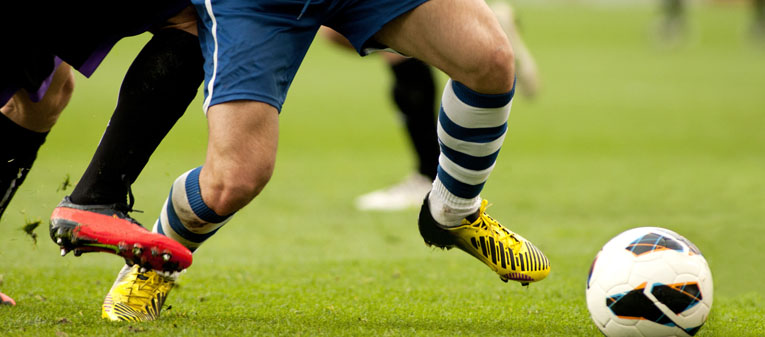
In general, the incidence of ACL injury is higher in people who participate in high-risk sports, such as basketball, football, and skiing. Approximately 50 percent of ACL injuries occur in combination with damage to the meniscus, articular cartilage, or other ligaments. Additionally, patients may have bruises of the bone beneath the cartilage surface.
It is estimated that 70 percent of ACL injuries occur through non-contact mechanisms, whilst the remaining 30 percent result from direct contact with another player or object. Immediately after the injury, patients usually experience pain and swelling and the knee feels unstable. Within a few hours after a new ACL injury, patients often have a large amount of knee swelling, a loss of full range of motion, pain or tenderness along the joint line and discomfort while walking.
The goal of the ACL reconstruction surgery is to prevent instability and restore the function of the torn ligament, creating a stable knee. This allows the patient to start playing sports again.
There are certain factors that the patient must consider when deciding for or against ACL surgery. Active adult patients involved in sports or jobs that require pivoting, turning or hard-cutting as well as heavy manual work are encouraged to consider having ACL reconstruction. Activity (NOT age) should determine if surgical intervention should be considered.
In young children or adolescents with ACL tears, early ACL reconstruction creates a possible risk of growth plate injury, leading to bone growth problems. We can delay ACL surgery until the child is closer to skeletal maturity or we may modify ACL surgery technique to decrease the risk of growth plate injury.
Before ACL reconstruction, the patient is usually sent to physiotherapy. Patients who have a stiff, swollen knee lacking full range of motion at the time of ACL surgery may have significant problems regaining motion after surgery. It is also recommended that some ligament injuries be braced and allowed to heal prior to ACL surgery (rare).
ACL reconstruction is usually performed under general anaesthesia. The surgery usually begins with an examination of the patient's knee while the patient is relaxed due to the effects of anaesthesia. This final examination is used to verify that the ACL is torn and to check for looseness of other knee ligaments that may need to be repaired during surgery or addressed postoperatively. Then, the selected tendon is harvested (for an autograft) or thawed (for an allograft) and the graft is prepared to the correct size for the patient. ACL tears are not usually repaired using suture to sew it back together, because repaired ACLs have generally been shown to fail over time.
The grafts commonly used to replace the ACL include:
- Patellar tendon autograft
- Hamstring tendon autograft
- Quadriceps tendon autograft
- Allograft (taken from a cadaver): patellar tendon, Achilles tendon and hamstring tendon
After the graft has been prepared, we place an arthroscope into the joint. Small incisions called portals are made in the front of the knee to insert the arthroscope along with the instruments. Following this, we then examine the condition of the knee. Meniscus and cartilage injuries are trimmed or repaired and the torn ACL stump is then removed.
In the most common ACL reconstruction technique, bone tunnels are drilled into the tibia and the femur to place the ACL graft in almost the same position as the torn ACL. The sutures of the graft are placed through the eye of the needle and the graft is pulled into position up through the tibial tunnel and then up into the femoral tunnel. The graft is held under tension as it is fixed in place using interference screws or posts. The devices used to hold the graft in place are generally not removed. Before the surgery is complete, the skin is closed and dressings are applied.
As with any surgery, there are risks associated with ACL reconstruction. These occur infrequently and are minor and treatable. Potential postoperative problems with ACL reconstruction include:
- Infection
- Bleeding
- Numbness
- Blood clot
- Instability (due to rupture or stretching of the reconstructed ligament)
- Stiffness
- Extensor mechanism failure (rupture of the patellar tendon or patella fracture may occur due to weakening at the site of graft harvest.)
- Growth plate injury in young children or adolescents
- Anterior knee pain (especially after patellar tendon autograft ACL reconstruction)
Physiotherapy is a crucial part of successful ACL surgery, with exercises beginning immediately after the surgery. Much of the success following ACL reconstructive surgery depends on the patient's dedication to a well planned physiotherapy programme.
In the first 10 days after surgery, the wound is kept clean and dry, and early emphasis is placed on regaining the ability to fully straighten the knee and restore quadriceps control. The knee is iced regularly to reduce swelling and pain. Weight-bearing status is also determined by the patient’s condition, including other injuries addressed at the time of surgery.
The goals for rehabilitation of ACL reconstruction include reducing knee swelling, maintaining mobility of the patella (to prevent anterior knee pain problems), regaining full range of motion of the knee, as well as strengthening the quadriceps and hamstring muscles. The patient may return to sports when there is no longer pain or swelling and when full knee range of motion has been achieved. Also, one’s muscle strength, endurance and functional use of the leg should be fully restored before returning to playing sports. Usually, it takes 5 to 6 months after surgery to return to full-contact sports.


