

About your knee
The knee is the largest joint in the human body, and healthy knees are required to perform most everyday activities.
The knee is made up of the lower end of the femur, the upper end of the tibia, and the kneecap (patella). The ends of these three bones, where they touch each other, are covered with articular cartilage that protects the bone ends and allows them to move smoothly.
The medial and lateral meniscus are located between the femur and tibia. These C-shaped wedges work as shock absorbers between two bones. Several ligaments hold the femur and tibia together and provide stability. The long thigh muscles, quadriceps muscle and biceps muscle, give the knee strength. All remaining surfaces of the knee are covered by the synovial membrane. This thin lining releases fluid that lubricate the cartilage, eliminating almost all friction in a healthy knee.
Various diseases or injuries can disrupt the cooperative system among them, resulting in pain, muscle weakness, or reduced function.

The knee is a major weight-bearing joint that is held together by muscles, ligaments and soft tissue. Cartilage can be found inside the joint and provides shock absorption which comes into play whenever we walk, run, lift, climb stairs and so on.
The femur and the tibia come together to form a hinge with the patella in front of them, which provides protection for the joint, the patella moves in a sliding action up and down in a groove in the femur. This groove is called the femoral groove and the sliding occurs whenever we flex or extend our knees.
Ligaments assure that the components of the knee are held together and kept stable. The medial collateral ligament (MCL) and lateral collateral ligament (LCL) limit sideways motion of the knee. The anterior and posterior cruciate ligaments (ACL and PCL) limit forward motion of the knee bones, thus keeping them stable.
Each knee has two cartilage structures called menisci, which sit between the femur and the tibia. These act as shock absorbers. The menisci are one of two types of cartilage in the knee. The second type, articular cartilage, is a smooth material which covers the end of the femur, the femoral groove, top of the tibia and the underside of the patella. These enable the knee and its bones to move smoothly.
Tendons connect muscle to the knee. The quadriceps muscles on the front of the femur are connected to the top of the patella by the quadriceps tendon, which covers the patella and becomes the patellar tendon. The patellar tendon then attaches to the front of the proximal tibia.
The hamstring muscles in the back of the leg attach to the distal femur at the back of the knee. The quadriceps muscles straighten the knee while the hamstring muscles provide the knee’s bending motion.
Knee pain can be caused by injuries, mechanical problems, types of arthritis and other problems.
A knee injury can affect any of the ligaments, tendons or fluid-filled sacs (bursae) that surround your knee joint as well as the bones, cartilage and ligaments that form the joint itself. Some of the more common knee injuries include ACL injury, fractures, torn meniscus, knee bursitis, and patellar tendinitis.
Also, some examples of mechanical problems that can cause knee pain include loose body, Iliotibial band syndrome (this occurs when the tough band of tissue that extends from the outside of your hip to the outside of your knee becomes so tight that it rubs against the outer portion of your femur), dislocated patella and hip or foot pain (Altered gait due to hip and foot pain can place more stress on your knee joint).
In addition, arthritis can cause knee pain. The varieties most likely to affect the knee include osteoarthritis, rheumatoid arthritis, gout, pseudo-gout (this is caused by calcium-containing crystals that develop in the joint fluid - knees are the most common joint affected by pseudo-gout), and septic arthritis.
Additionally, patellofemoral pain syndrome (pain arising between your patella and the femur) is common in young adult athletes.
A number of factors can increase your risk of having knee problems, including excess weight, lack of muscle flexibility or strength, certain sports (e.g. alpine skiing with its rigid ski boots or basketball's jumps and pivots) and previous injury. Having a previous knee injury makes it more likely that you'll injure your knee again.
A swollen knee usually occurs when excess fluid accumulates in or around the knee joint. This condition can be referred to as an effusion in the knee joint.
A swollen knee may be the result of trauma, overuse injuries, or an underlying disease or condition. To determine the cause of the swelling, we might need to obtain a sample of the fluid to test for infection, disease or injury. Removing some of the fluid also helps reduce the pain and stiffness associated with the swelling.
Many types of problems, ranging from injuries (torn ligament particularly ACL, cartilage (meniscus) tear, irritation from overuse, and broken bones), to diseases and conditions (osteoarthritis, rheumatoid arthritis, infection, gout, pseudo-gout, bursitis and tumours) can cause a swollen knee.
Clicking knees are often caused by injuries or stress to the knee or leg. Over time, or after an injury, you may notice that your knee begins to click when you move. Sometimes this condition is accompanied by pain, but normally there are no other symptoms besides the noise. This is typically caused by some portion of the knee not sitting in the proper position, so different parts of the leg are being used and stressed more than normal.
There are many conditions that can cause the knee to start clicking, most of which are harmless. Some of the common causes are: unnecessary tissue or plica (Some people develop additional unnecessary tissue or plica around the knee. This tissue can then become trapped between parts of the joint, which will cause a clicking noise when you move), runner's knee (This is caused when the patella is out of line and not tracking properly along the femur. You may also suffer runner's knee if the band which stabilises the patella or your quad muscles are overworked), damage of the meniscus, and arthritis.
If the clicking is causing you pain, or making it difficult to move, then it is important to seek medical attention to determine whether you are suffering from an injury that could be causing damage to the knee.
Loss of movement, such as, knee stiffness, can happen on its own or because of injury or surgery. The most common cause of loss of motion in the knee is arthritis. The most common form of arthritis is osteoarthritis but it can be from rheumatoid arthritis or post-traumatic arthritis as well.
Other inflammatory conditions which cause knee swelling such as gout, psoriatic arthritis and PVNS (pigmented villonodular synovitis) can cause loss of knee movement too. The stiffness from osteoarthritis tends to develop slowly over time. It gradually worsens as the pain in the knee increases and you might develop a fixed flexion deformity due to scar tissue forming around the knee. Most people notice this symptom because they have trouble putting their shoes or socks on, or have difficulty standing from a chair.
Also, a meniscal tear can cause your knee to be stiff but this is not the usual presentation for this condition. Joint cartilage injuries can cause knee stiffness as well.
Giving way is when the knee suddenly 'gives out', causing you to stumble or fall.
There are two main types of giving way of the knee. One is an intermittent giving way associated with sudden sharp pains in the knee, another is a feeling of wobbliness in the knee with regular giving way. Intermittent painful giving way, especially if associated with painful clicking, is often due to a meniscal cartilage tear, where torn flaps of cartilage get caught between the knee bones.
A knee that feels generally wobbly may have ligamentous instability, due to a torn ligament such as the ACL. Other potential causes of giving way are unstable flaps of articular cartilage or loose bodies (loose pieces of cartilage and/or bone floating around within the knee).
The major types of arthritis that affect the knee are osteoarthritis, rheumatoid arthritis, spondyloarthritis, and psoriatic arthritis.
Of these, osteoarthritis is the most common form of arthritis in the knee. It is a degenerative arthritis that occurs most often in people 50 - 60 years of age and older. In osteoarthritis, the cartilage in the knee joint gradually wears away. As the cartilage wears away, it becomes frayed and rough, and the protective space between the bones decreases. This can also result in painful bone spurs. Osteoarthritis develops slowly and the pain it causes worsens over time. As with other arthritic conditions, initial treatment of osteoarthritis of the knee is nonsurgical. We may recommend a range of treatment options, such as physical therapy, losing weight, using assistive devices (e.g. canes), or medications. We may recommend surgery (e.g. total or partial knee replacement) if your pain from osteoarthritis causes disability and is not relieved with nonsurgical treatment.
Rheumatoid arthritis is a chronic disease that attacks multiple joints throughout the body, including the knee joint. It usually affects the same joint on both sides of the body (symmetrical). In rheumatoid arthritis, the synovial membrane that covers the knee joint begins to swell, this leads to knee pain and stiffness. Rheumatoid arthritis is an autoimmune disease. The immune system attacks its own tissues, in other words, the immune system damages normal tissue (such as cartilage and ligaments) and softens the bone.
Spondyloarthritis is a name for a family of inflammatory rheumatic diseases that cause arthritis, which attacks the spine and, in some people, the joints including the knee. It can also involve the skin, intestines and eyes. Though the main symptom in most patients is low back pain, in a minority of patients the major symptom is pain and swelling in the knee. People in their teens and 20s, particularly males, are affected most often. Family members of those with spondyloarthritis are at higher risk.
Psoriatic arthritis is a type of inflammation that occurs in about 15 percent of patients who have a skin rash called psoriasis. In some people, it can affect the large joints, especially those of the lower extremities including the knee. Early diagnosis is important to avoid damage to joints. Psoriatic arthritis can occur in people without skin psoriasis, particularly in those who have relatives with psoriasis. Psoriatic arthritis usually appears in people between the ages of 30 to 50, but can begin as early as childhood. Men and women are equally at risk.
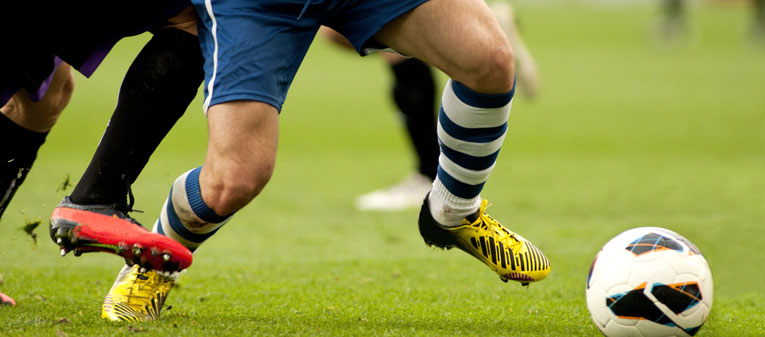
An anterior cruciate ligament sprain or tear is one of the most common knee injuries. Athletes who participate in high demand sports like soccer, football, and basketball are more likely to injure their ACL. If you have injured your ACL, you may require surgery to regain full function of your knee. This will depend on several factors, such as the severity of your injury and your activity level.
When you injure your ACL, you might hear a noise and you may feel your knee give out from under you. Other typical symptoms include pain with swelling, loss of full range of motion, tenderness along the joint line, and discomfort while walking.
Treatment for an ACL tear will vary depending upon the patient's individual needs. For example, the young athlete involved in agility sports will most likely require surgery (rebuilding the ligament arthroscopically) to safely return to sports. The less active, usually older, individual may be able to return to a quieter lifestyle without surgery.
PCL is located in the back of the knee. It is one of several ligaments that connect the femur to the tibia. PCL keeps the tibia from moving backwards too far. An injury to the posterior cruciate ligament requires a powerful force. A common cause of injury is a bent knee hitting a dashboard in a car accident or a football player falling on a knee that is bent.
Injuries to PCL are often subtle and more difficult to evaluate than other ligament injuries in the knee. PCL tears tend to be partial tears with the potential to heal on their own. People who have injured just their PCL are usually able to return to sports without knee stability problems.
Basically, patients may be recommended simple, nonsurgical options. We may recommend surgery if you have combined injuries. For example, if you have dislocated your knee and torn multiple ligaments including PCL, surgery is almost always necessary.
Your knee ligaments connect your femur to your lower leg bones. Knee ligament sprains or tears are a common sports injury. Athletes who participate in direct contact sports like football or soccer are more likely to injure their collateral ligaments.
The medial collateral ligament (MCL) connects the femur to the tibia. The lateral collateral ligament (LCL) connects the femur to the fibula. The collateral ligaments control the sideways motion of your knee and brace it against unusual movement.
Any direct contact to the knee or hard muscle contraction, such as changing direction rapidly while running, can injure a knee ligament.
Most isolated collateral ligament injuries can be successfully treated without surgery. If the collateral ligament is torn in such a way that it cannot heal, or is associated with other ligament injuries such as ACL, we may suggest surgery to repair it.
Three bones (femur, tibia and patella) meet to form the knee joint.
Bones are connected to other bones by ligaments. There are four primary ligaments in your knee. Collateral ligaments are found on the sides of the knee. The medial collateral ligament (MCL) is on the inside and the lateral collateral ligament (LCL) is on the outside. They control the sideways motion of the knee.
Cruciate ligaments are found inside the knee. They cross each other to form an "X" with the anterior cruciate ligament in front and the posterior cruciate ligament in back. The cruciate ligaments control the back and forth motion of the knee. The knee joint relies just on these ligaments for stability.
Direct contact to the knee or hard muscle contraction can injure a knee ligament. It is possible to injure two or more ligaments at the same time. These multiple injuries can have serious complications. The MCL is injured more often than the LCL.
In contrast to treatment for single ligament tears, surgery for combined ligament tears is often performed soon after the injury. More than one operation may be required when treating multiple ligament injuries.
Meniscus tear is common knee injury. Athletes, particularly those who play contact sports, are at risk of meniscus tears. However, anyone at any age can tear their meniscus.
Meniscus are two wedge-shaped pieces of cartilage working as shock absorbers between your femur and tibia. Meniscus tears are noted by how they look, as well as where the tear occurs in the meniscus. Common tears include bucket handle, flap, and radial. Sports-related meniscus tears often occur along with other knee injuries, such as ACL tears.
You might feel a pop when you tear a meniscus. Most people can still walk on their injured knee. Over several days, your knee will gradually become stiffer and swollen.
How we treat your tear will depend on the type of tear you have, its size, and location. Along with the type of tear you have, your age, activity level, and any related injuries will also factor into your treatment plan. If your symptoms persist with nonsurgical treatment, we may suggest arthroscopic surgery.
The OCD is a lesion of the cartilage and bone due to necrosis and loss of continuity of the underlying bone.
The cause of this lesion is not known. There is a history of trauma to the knee in some patients. The most common site is the medial femoral condyle (80%). Other sites are the lateral femoral condyle (15%), and the patella (5%).
Males are 3 times more likely to be affected than females, and it most often occurs in children. Frequently, it presents without a traumatic event as a painful knee with vague, poorly localised symptoms.
The pain is usually only with the weight-bearing and does not develop at rest. Sometimes locking or clicking will also be present with knee motion and walking.
The OCD lesions have a greater chance of separating from the surrounding bone and cartilage, and can even detach and float around inside the joint. In these cases, surgery may be necessary.
This occurs when a segment of femoral or tibial bone loses its blood supply and begins to die.
More than 3 times as many women as men are affected; most are over the age of 60 years. The exact cause of AVN (another name is the osteonecrosis of the knee) is not yet known. One theory is that a stress fracture, combined with a specific activity or trauma, results in an altered blood supply to the bone.
AVN of the knee is also associated with certain conditions and treatments, such as obesity, lupus, and steroid therapy. Steroid-induced osteonecrosis frequently affects multiple joints and is usually seen in young patients.
Regardless of the cause, if the disease is not identified and treated early, it can develop into severe osteoarthritis.
Osgood-Schlatter disease is a painful knee condition that affects adolescents.
It is thought that the patellar tendon attaching the quadriceps muscle to the knee joint becomes tighter, creating a strain on the growing bone, particularly with physical activities that involve contraction of the quadriceps (e.g. soccer) during a growth spurt.
Boys are affected more than girls, although this could be due to differing activity patterns.
Osgood-Schlatter disease usually resolves by itself with a period of activity modification, ice, pain-relieving medications, stretching and physiotherapy. Only in extreme cases and only once growth has ended, surgery could be considered.
PVNS is a condition that causes the synovium to thicken and overgrow. The mass or tumour that results from this overgrowth is not cancerous and does not spread to other areas of the body.
PVNS is a benign disease, however, it is a progressive disease that slowly worsens and can lead to bone damage and arthritis. PVNS usually affects the knee although it can affect other joints as well (e.g. the hip).
In most cases, surgery is needed to remove the damaged joint lining and the mass. The condition can affect people of all ages, but it occurs most often in young adults in their 30s.
There are two forms of PVNS. When the tumour involves the tendons that support the joint, or occurs in just one area of the joint, it is called localised PVNS, which usually responds well to treatment. When the condition is more widespread and involves an entire joint, it is called diffuse PVNS, which tends to be more destructive and is more difficult to treat.
Synovial chondromatosis is a rare, benign condition that involves the synovium. Although this type of tumor does not spread to other parts of the body, it can cause severe damage to the joint and lead to osteoarthritis.
In synovial chondromatosis, the synovium grows abnormally and produces nodules made of cartilage in the joint. These nodules may break off from the synovium and become loose inside the joint, including the hip. In severe cases, the loose bodies may grow large enough to occupy the entire joint space or penetrate into adjacent tissues.
Synovial chondromatosis most often occurs in the knee, followed by the hip. In most cases, only one joint in the body is affected.
Treatment for synovial chondromatosis involves surgery to remove the loose bodies of cartilage. This can be done with an open surgical procedure or knee or hip arthroscopy.
The patella runs in the trochlea along the front of the knee. It is kept in place by the bony contours of this groove, as well as ligaments that keep the patella from moving side to side. The patella can be forced out of its groove during a knee injury, causing the knee to become dislocated.
In some cases, the patella can slip partially out of place and then quickly reduced. There is often intense pain and swelling associated with the patella dislocation. If the instability becomes chronic, the patella may frequently slide out of place. These episodes can become less painful but more frequent over time.
After a patella dislocation, the lower extremity may be placed in an immobiliser brace for a period of time to allow the patella to rest and heal. Most people who undergo rehabilitation appropriately after a first patella dislocation tend to not have recurrent problems.
In the less common case of repetitive patellar dislocations, surgery can be done to stabilise the patella and prevent it from sliding out.
-

- Assessment
Hip and knee joints connect your bones and allow the movement of your lower extremity. Pain or discomfort in these joints can be caused by injury.
-

- Treatments
Depending on the cause of your pain, the solution might be a set of exercises, pain relief medication, minor/major surgery, or a combination of these.
-

- Make an appointment
If you have any further questions or wish to make an appointment, please do not hesitate to get in touch via telephone or email on our contacts page.
-

- Testimonials

Dame Jessica Ennis Hill
Olympic Gold Medallist

Christian Knightly
Hip Replacement Patient - 100 mile cycle charity challenge
Thomas Horton
Hip Arthroscopy Patient - Football

